Information

Information

TowardPi believes in long-term value and adheres to a democratic company management model, with the spirit of scientists at its core
Your location:
Homepage
/
News
2024-04-23

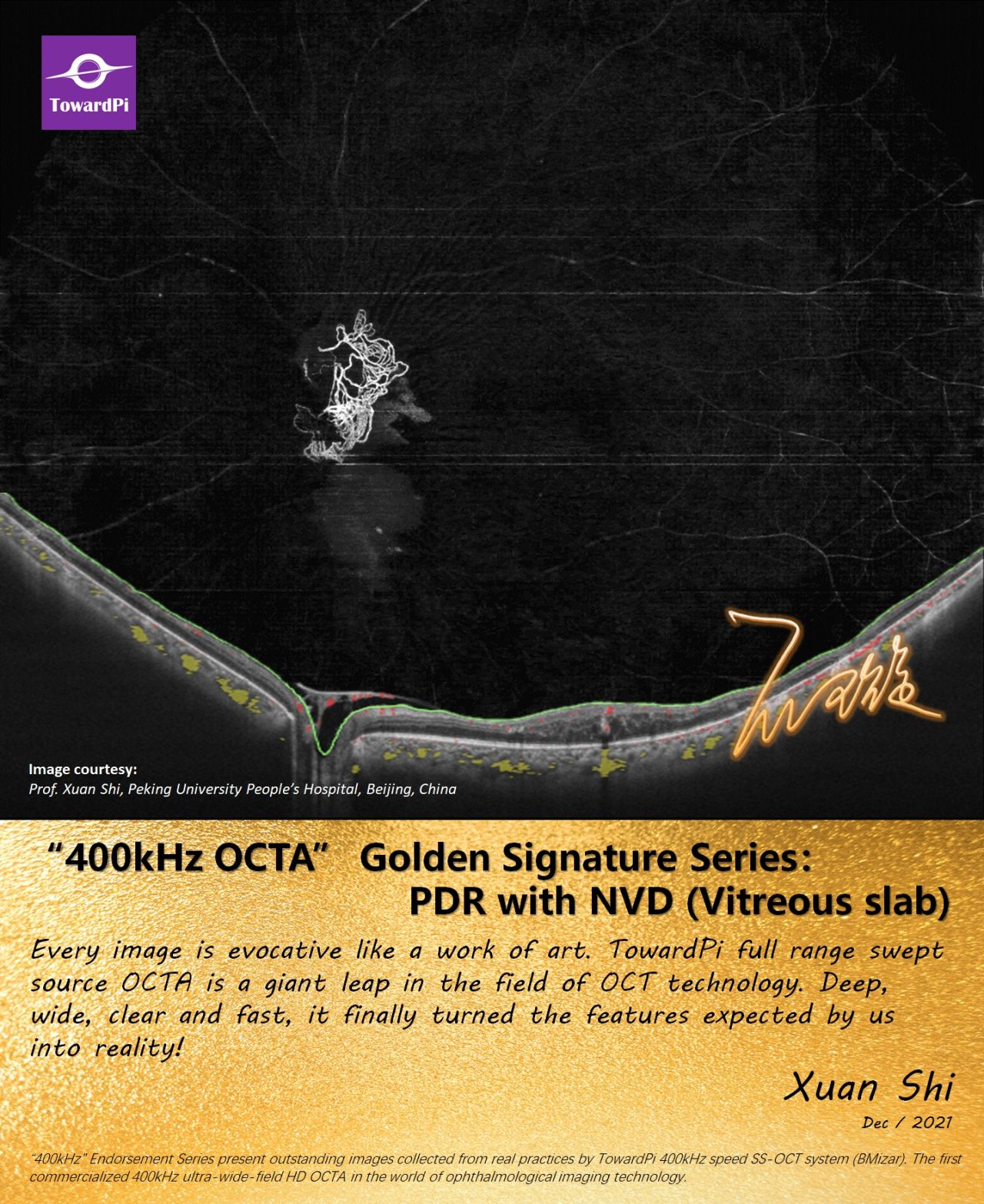
Proliferative Diabetic Retinopathy, PDR with NVD (Vitreous slab), OCTA single scan

Proliferative Diabetic Retinopathy, PDR with NVD (Vitreous slab), OCTA single scan (1536 x 1280) ‘Every image is evocative like a work of art. TowardPi's full -range swept-source OCTA is a giant leap in the field of OCT technology. Deep, wide, clear and fast, it finally turned the features expected by us into reality!’
2024-04-22

Posterior Vortex Veins in Healthy Eyes Captured by Ultra-widefield OCTA

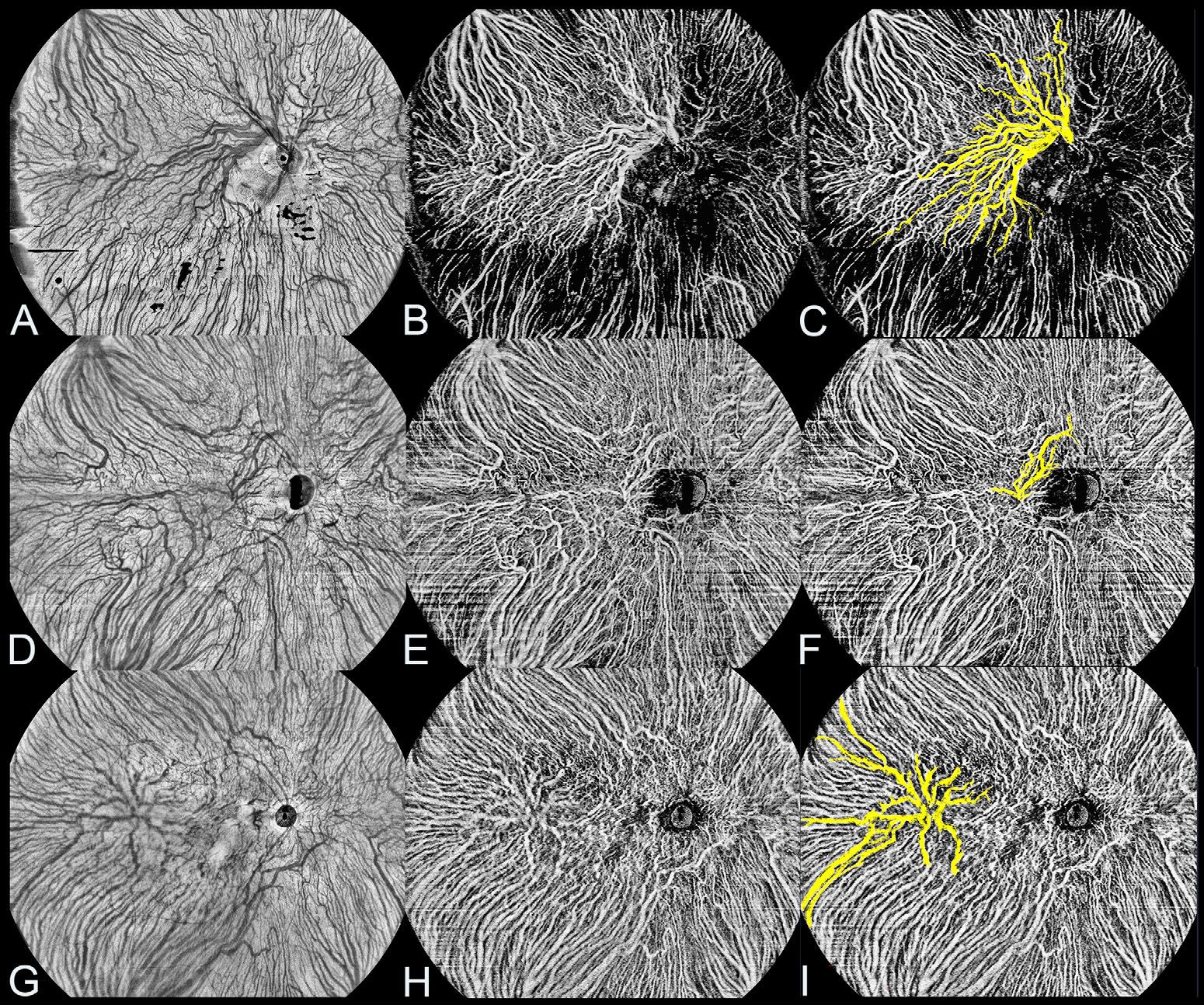
Perfectly consistent with ICG New research published in the Investigative Ophthalmology & Visual Science investigated the incidence and characteristics of posterior vortex veins (PVVs) in healthy eyes and explored their relationship with age and refractive status by using ultra-widefield OCTA (UWF-OCTA). This study confirmed PVVs' presence in healthy eyes (16.1%, 82/510) and classified PVVs into three types based on the location of the vortex vein exit from the eyes: those around the optic disc (fig.1 C), those in the macula area (fig.1 F), and those in other areas of the posterior pole (fig.1 I). Interestingly, this study revealed a predominant presence of PVVs around the optic disc (fig.2), followed by other areas.
2024-04-12

Corolla in the Fundus, Ultra-widefield OCTA of Takayasu Arteritis

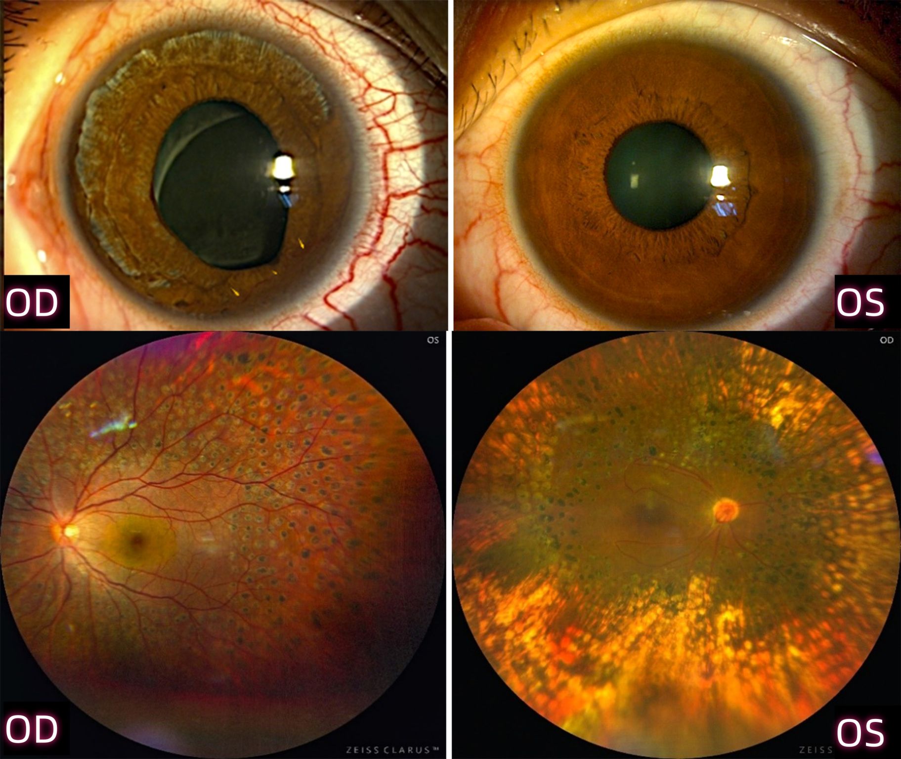
29-year-old female presented with abnormally increased C-reactive protein and erythrocyte sedimentation rate was diagnosed as Takayasu arteritis complicated with severe retinopathy. The scleral vessels in her right eye were observed highly dilated with iris stroma atrophy and irregular pupil as shown in the picture. Both eyes underwent pan-retinal photocoagulation (fig.1).
2024-04-10

A Wireless Battery-free Eye Modulation Patch for High Myopia Therapy

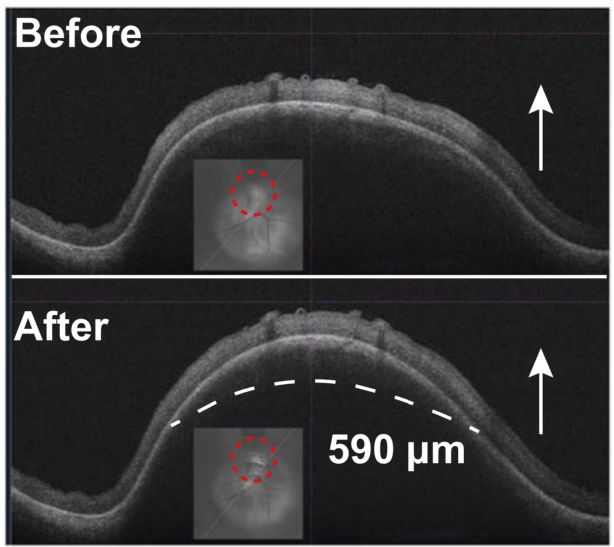
The new study recently published in (IF=16.6) presented a battery-free eye modulation patch designed to correct high myopia and prevent relapse. Positioned on the sclera near the optic nerve, corresponding to the macular area, the patch uses piezoelectric transduction to convert external ultrasound into electrical energy, and then controls the device’s components. The micro-actuator creates gas bubbles, causing a membrane to expand and retract the sclera behind the macula, effectively shortening AXL (axial length) for vision correction. The microneedle array delivers riboflavin to the posterior sclera, while μ-LEDs induce scleral collagen cross-linking (SCXL), strengthening the sclera against high myopia-induced relaxation.
2024-04-08

Introducing TowardPi's OCT to Singapore Deputy Prime Minister, Mr. Heng Swee Keat

We were honored to introduce TowardPi's self-developed OCT and other devices to Singapore Deputy Prime Minister, Mr. Heng Swee Keat. DPM Heng highly appreciated the rapid progress of our high-end ophthalmic technology. Meanwhile, we would also like to express our gratitude to our incubation platforms, Tsinghua University and TusStar for their strong support in this visit.
2024-04-02

IRVAN Syndrome Revealed by UWF Full-range OCTA

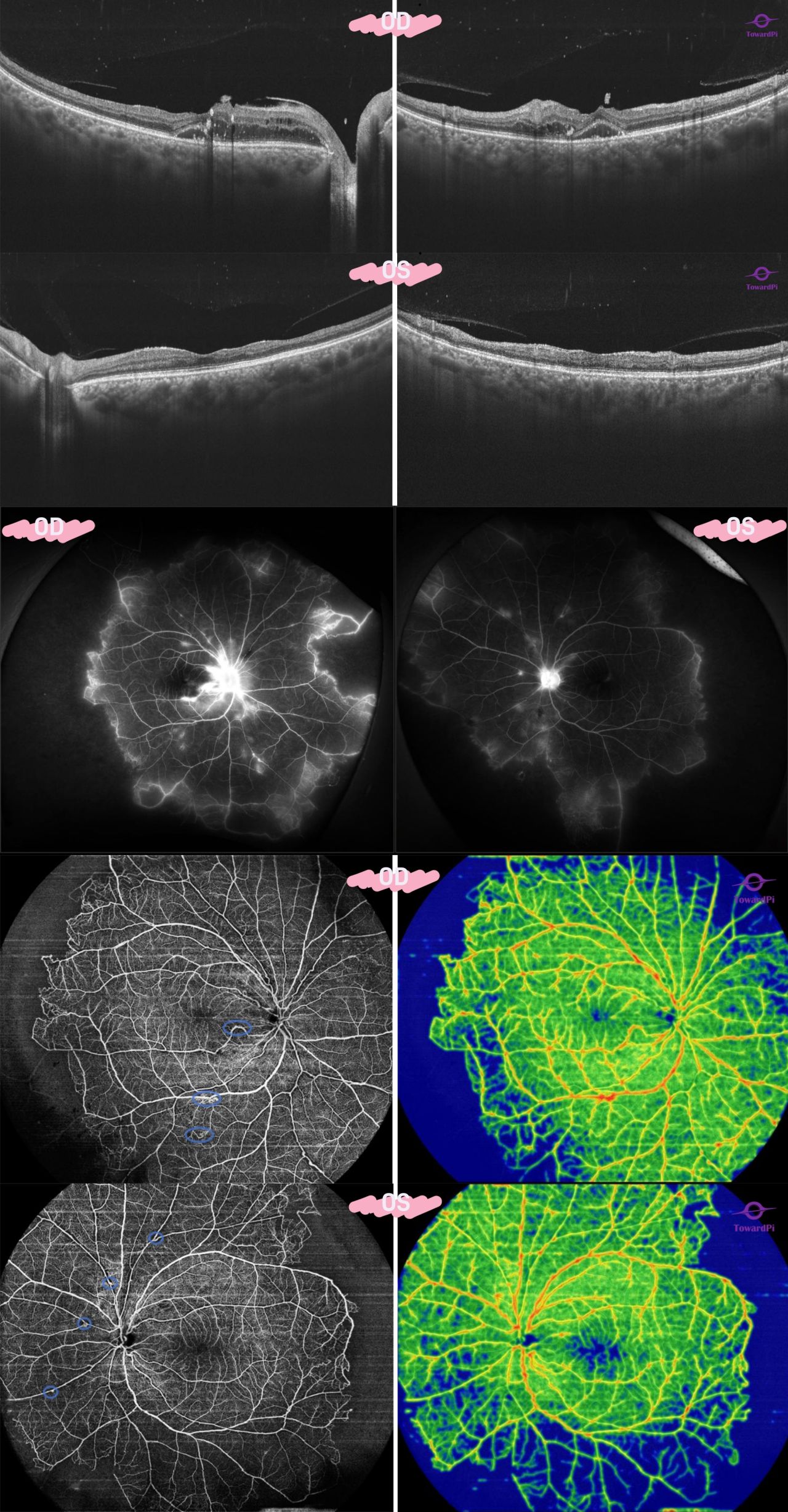
A 52-year-old female presented with hard exudation and intraretinal cystoid edema in her right eye and retinal atrophy in her left eye as shown in the high-resolution B-scan images. UWF full range OCTA revealed typical non-perfusion peripheral temporal area in her both eyes and aneurysmal ectasia with tortuous small vessels around the optic disks (blue circle). IRVAN syndrome (idiopathic retinal vasculitis,aneurysms and neuro-retinitis syndrome) was considered as the diagnosis
2024-03-27

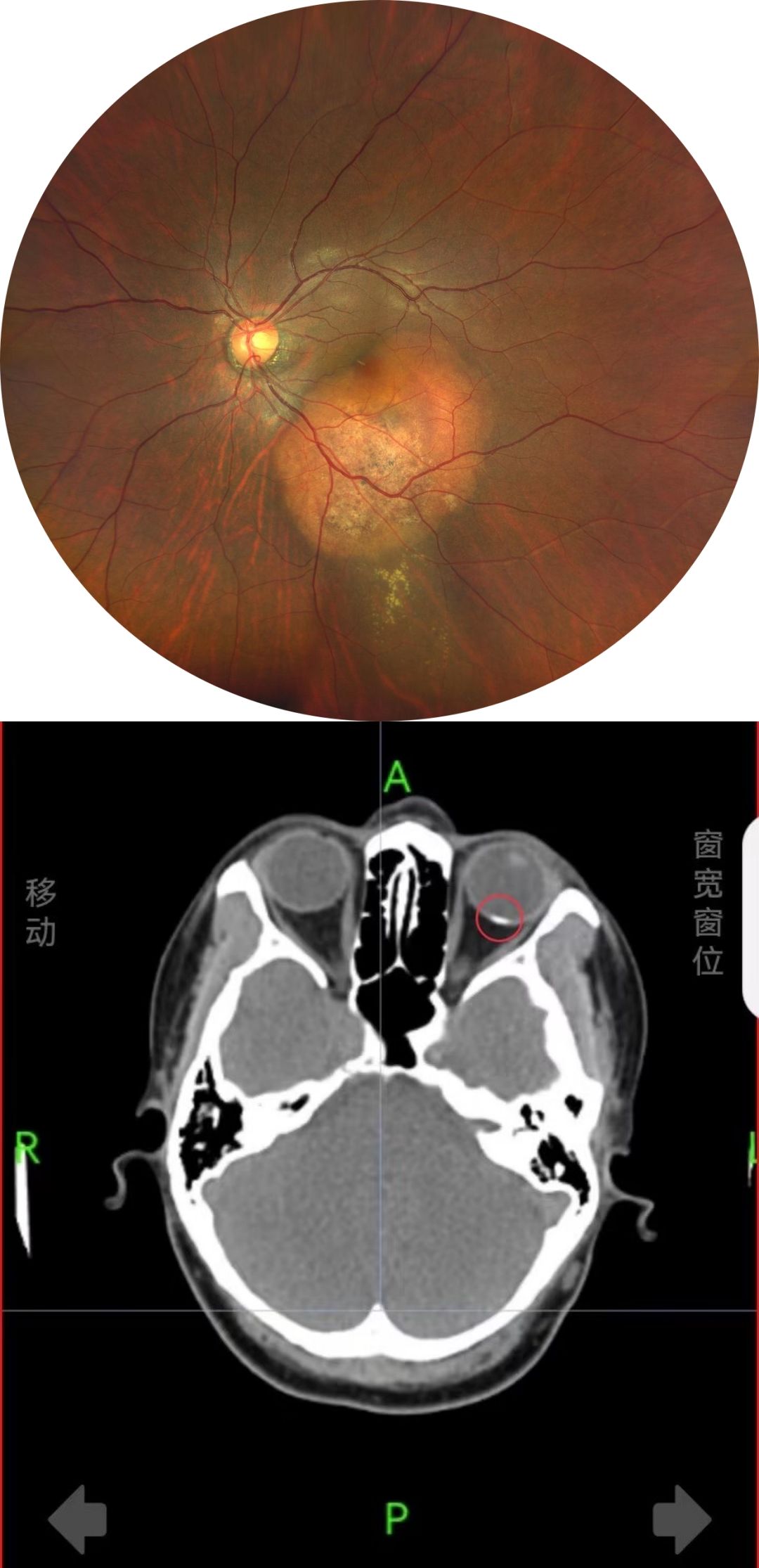
Choroidal Osteoma with Secondary CNV Captured with Full-range SS-OCTA

A 37-year-old female presented with orange irregular oval intraocular tumor at the posterior pole in the left eye as the color fundus shown. CT revealed an arc-shaped high radiodense lesion consistent with bone density at the posterior pole of eyeball (fig.1).
2024-03-22

Virus Keratohelcosis with Secondary Corneal Neovascularization Captured by AS-OCTA

Anterior segment OCTA revealed that large corneal neovascular membrane invaded from the nasal side and communicated with the large vessels of the nasal sclera (fig. a). Flow area of the neovascularization could be quantified with built-in quantification tool (fig. b). The entire cornea was thickened with diffuse edema as shown in the en face image and high-resolution B-scan (fig. c&d).
2024-03-19

Dolphin in the Fundus

A 18-year-old male presented with an oval translucent cystic mass in the inferior nasal peripheral in his left eye as the color fundus (fig. A) shown. High-resolution swept-source OCTA revealed a 10.5 ×5mm "dolphin-shaped" epiretinal cystic hyperreflectivity on cross section image (fig. C) with a little blood flow signals detected in the "cystic wall" (fig. D). A diagnosis of congenital cyst of the vitreous was considered.
2024-03-19

Advances in SS-OCT and OCTA Published in the Advances in Ophthalmology Practice and Research

Recently "Advances in SS-OCT and OCTA”was published in the Advances in Ophthalmology Practice and Research. It provides a comprehensive review of swept-source OCT technology and its advances in many applications. The article was authored by the team of Prof. Xiaoyun Fang.
Previous page
1
2
16

© TowardPi (Beijing) Medical Technology Ltd     京ICP备2021011388号-1   Powered by www.300.cn     View business license