Information

Information

TowardPi believes in long-term value and adheres to a democratic company management model, with the spirit of scientists at its core
Your location:
Homepage
/
/
Gallery
2025-03-07

“Motorbike” in Anterior Chamber Captured by TowardPi Full-Range AS-OCT

A 70-year-old male experienced persistent, distending pain in his right eye 11 days ago without an obvious cause, accompanied by ipsilateral headache, photophobia, and tearing. His right eye was found to have no light perception, with high intraocular pressure (51 mmHg), conjunctival hyperemia, a dilated pupil, and corneal haze and edema. High-resolution panoramic anterior segment OCT (AS-OCT) clearly showed the lens dislocated into the anterior chamber with lens protein extravasation, and a membranous protrusion was visualized in the pupillary area. This patient was diagnosed with phacogenic glaucoma secondary to lens dislocation.
2025-02-24

Proliferative Diabetic Retinopathy Captured by 400kHz SS-OCTA

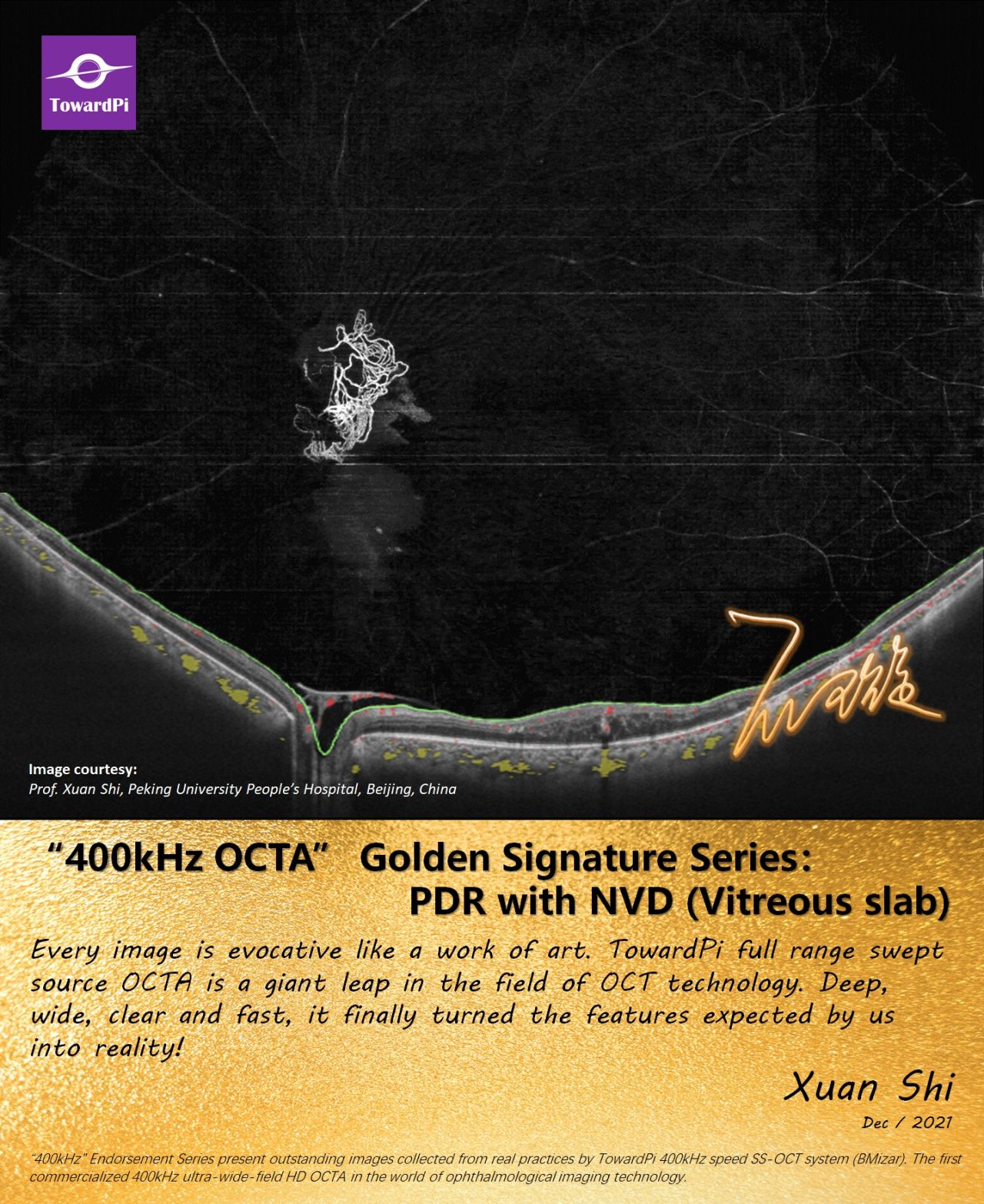
Proliferative Diabetic Retinopathy captured by 400kHz SS-OCTA. “TowardPi OCTA reveals the presence of retinal neovascularization which is the hallmark of the disease, allowing us to set an accurate diagnosis quickly (no dilation) without the need of interventional examinations (e.g . FFA) . Wide-field OCTA images allows the accurate assessment of extensive areas of retinal ischemia!” Golden Signature Series of TowardPi. Endorsement and courtesy: Dr. Ioannis Datseris, (President of Greek Vitreoretinal Society / Founder of OMMA Ophthalmic Institute Athens)  
2025-02-19

Resolve the Crisis of Choroidal Hemangioma with UWF Full Range SS-OCTA!

A 59-year-old male patient complained diminution of vision for recent 4 months. A round orange-red solitary mass superior nasal to optic disc was discovered by slim-lamp examination. UWF SS-OCT revealed significant elevated choroid corresponding to the solitary mass on SLO image together with irregular and disrupted RPE and outer retina, intraretinal edema and subretinal fluid under fovea (fig.1 OCT). Hyperreflective spots in vitreous could also be seen. On UWF SS-OCTA image of choriocapillaris slab, a dense vascular network in the tumor area was clearly visualized compared with the surrounding normal choriocapillaris (fig.1). UWF OCTA and quantifications of choroidal Haller’s and Sattler’s layer revealed the significantly thickened tumor and the medium-sized vessels inside the tumor with high vessel density (fig.2). This patient was diagnosed as circumscribed choroidal hemangioma. 3D UWF OCTA reconstructed the stereo structure of the fundus and the relationship between hemangioma and disc (gif). A 400kHz speed swept source OCTA (#BMizar, TowardPi Medical) was used in this case to capture ultrawide field 24mm x 20mm full range SS-OCTA images of choroidal hemangioma. OCTA of choroidal Haller’s and Sattler’s layer can be achieved with built-in algorithm together with comprehensive quantifications. 3D reconstruction of structures and vessels are available for all-sizes. Case courtesy of: Prof. Huiying Rao from Fujian Provincial Hospital. Editor: Dr. Chloe Li, TowardPi Medical See more: https://www.linkedin.com/posts/towardpi_bmizar-ophthalmology-ssoct-activity-7297517720621723648-1CwS?utm_source=share&utm_medium=member_desktop&rcm=ACoAACHzlMkBXV1jeYivCSt0EWunBEwGyFaWzXc 
2025-02-12

‘Ghost’ Vessels in Retinitis Pigmentosa Revealed by UWF SS-OCTA!

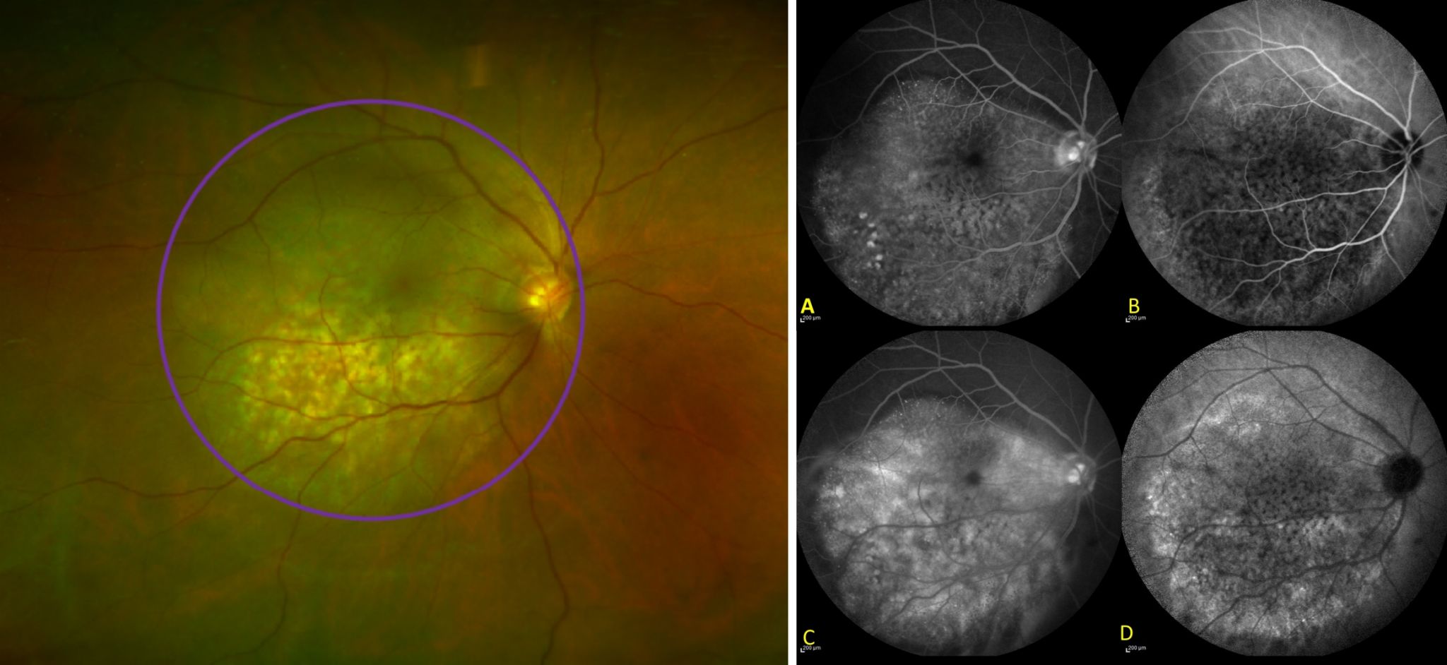
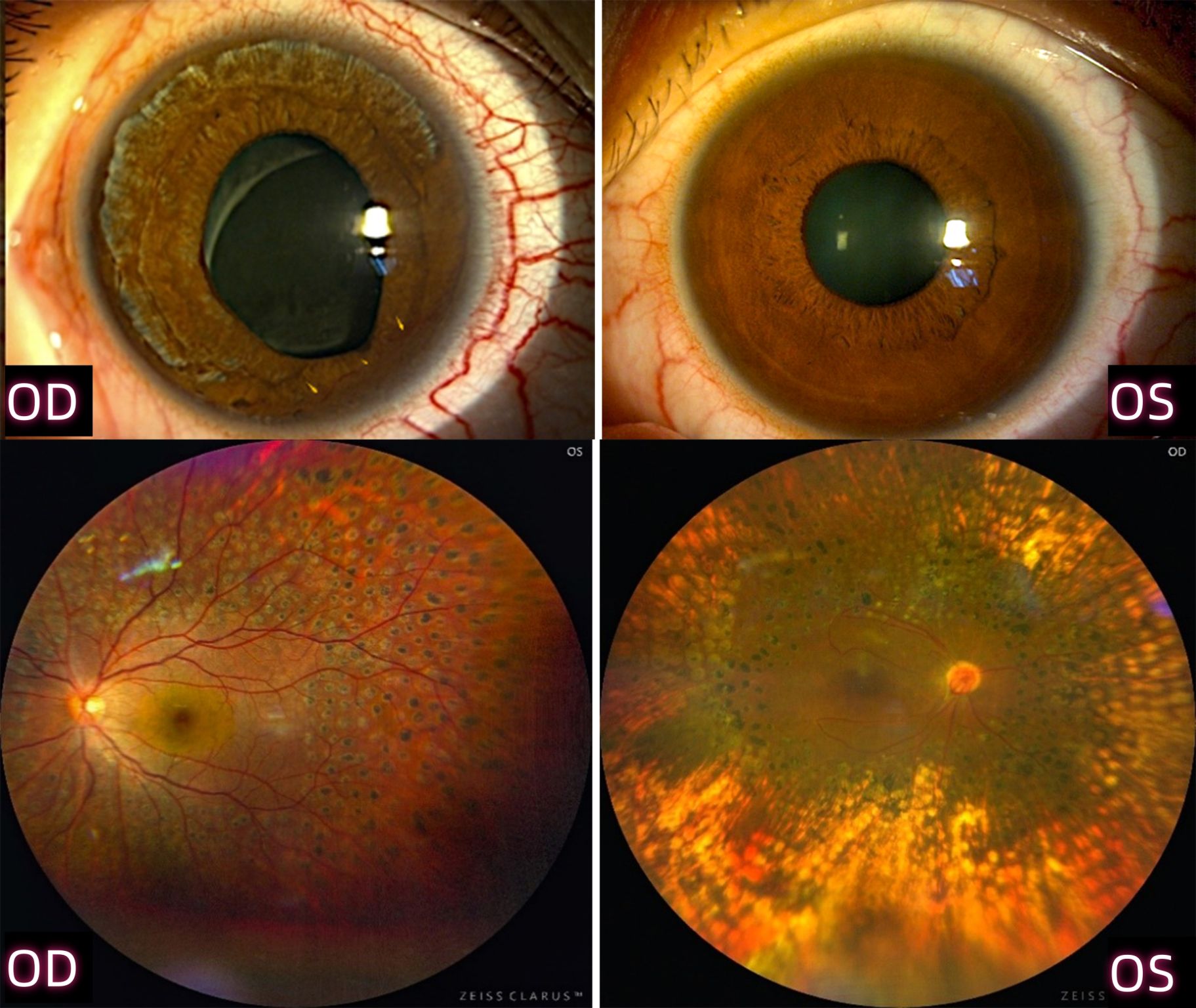
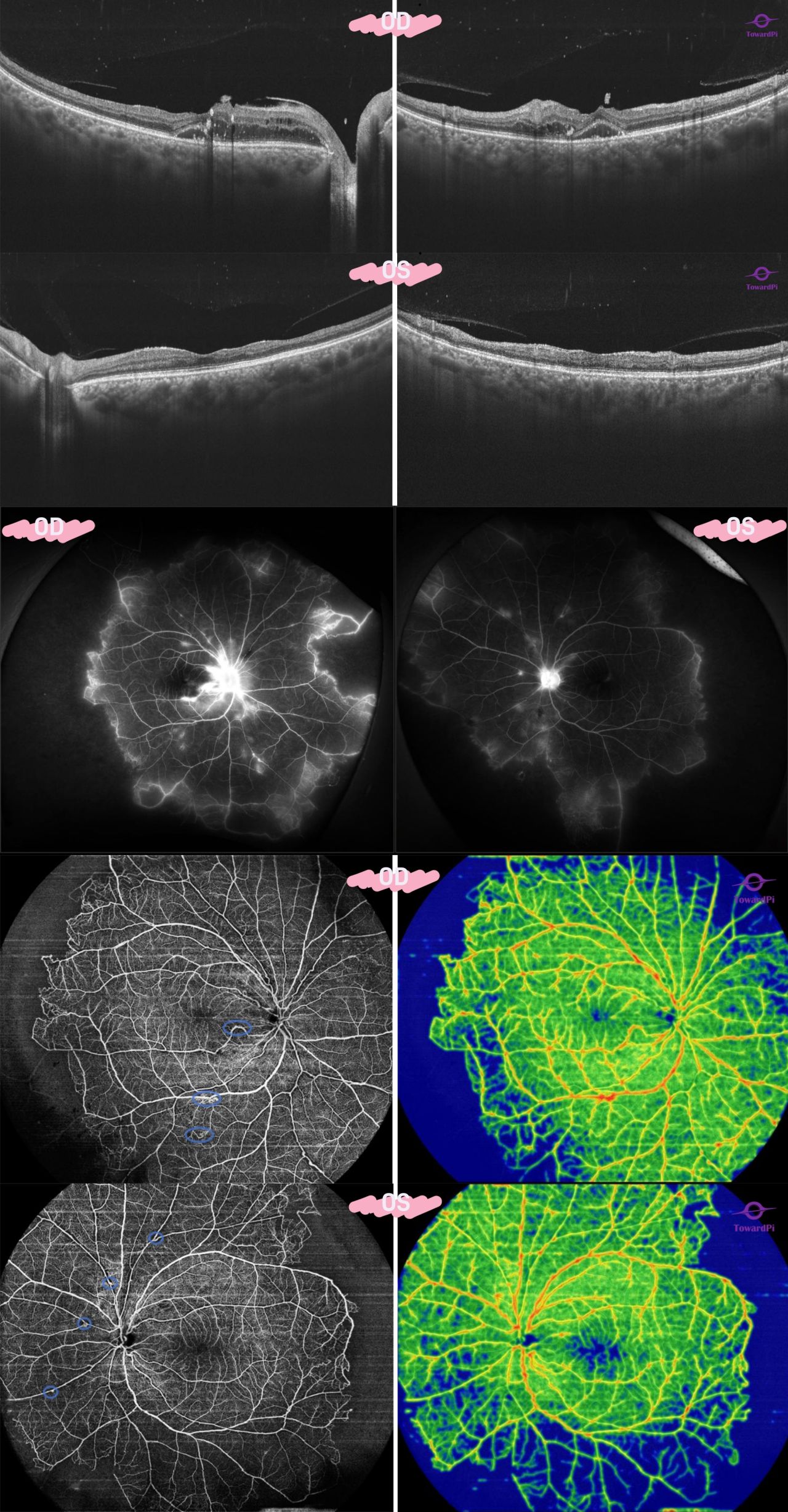
‘Ghost’ vessels in Retinitis pigmentosa revealed by UWF SS-OCTA! A 48-year-old male patient who had suffered from night blindness and decreasing peripheral vision for 40 years was diagnosed as retinitis pigmentosa (RP). Typical pigment deposits (fig.1 red arrow) in the mid periphery were shown on the color fundus images in his both eyes. Hyper-autofluorescent rings at post pole around macula were revealed by autofluorescence imaging in both eyes correlating with the relatively preserved photoreceptor layer at macula while peripheral hypo-autofluorescent (fig.1 green arrow)was observed. Ultrawide field 24mm B-scan images showed thinning of the photoreceptor layers with loss of the ellipsoid zone and remaining RPE outside of macula while atrophy retina and RPE or even absence of RPE and the outer nuclear layer in the mid periphery. Intraretinal hyperreflective spots in the periphery could also be observed (fig.1 orange square). On the UWF full range SS-OCTA images, typical vascular features of RP including vessel narrowing and sclerosis, significative reduction of retinal vessels in the mid periphery and nonperfusion periphery were clearly revealed, correlating to the severely reduced retinal vessel density (fig.2). In fact, the remaining vessel walls of some occluded retinal vessels could still be seen on the En face images of deep retinal vessel layer (fig.2 purple arrow) together with large amount of pigment deposits (fig.2 red arrow). A 400kHz speed swept source OCTA (#BMizar, TowardPi Medical) was used in this case to capture ultrawide field 24mm x 20mm full range SS-OCTA images of vascular abnormalities and retinal degeneration of RP. Precise segmentation algorithm and flow quantifications are available on the device for disease assessment. Case courtesy of: Dr. Jieqiong Chen, team of Prof. Xiaodong Sun from Shanghai General Hospital. Editor: Dr. Chloe Li, TowardPi Medical  
2025-02-07

Astonishing Auto-Segmentation of TowardPi Full-Range Swept-Source OCT/OCTA

Putting aside the exquisite choroid OCTA picture (above), the perfect accuracy of auto-segmentation alone is also astonishing (below). And this, is just one of the hundreds of standard functions from TowardPi full-range swept-source OCT/OCTA.  
2025-02-03

Asteroid Hyalosis with Retinal Detachment

Asteroid Hyalosis with retinal detachment. 3D (L) and Radial lines (R) captured by Yalkaid, a full-range swept-source OCT from TowardPi. Default setting (no further extended length or depth).
2025-01-29

Retinal Macroaneurysm revealed by 3D full range SS-OCTA!

Retinal Macroaneurysm revealed by 3D full range SS-OCTA! An 82-year-old female who had a history of arteriosclerosis complained vision loss in her right eye. The visual acuity was 20/50 in her right eye while 20/30 in her left eye. Intraocular pressure in both eyes was normal. Color fundus images discovered bilateral refractive media opacity and a spindle-shaped lesion nearby a small branch retinal artery inferotemporal to optic disc in her right eye (fig. 1 blue circle). High-resolution B-scan image revealed thickened retina with subretinal fluid (fig. 1 thickness map). An oval hyperreflective entirety was found located in the internal retina with relatively low internal reflection, corresponding to the spindle-shaped lesion on color fundus and SLO image (fig.1 blue arrow). 12mm x 12mm OCTA image clearly showed a retinal macroaneurysm with internal blood signals detected on the flow-overlaid B-scan image (fig. 2). It was more obvious with higher flow density on the vessel density map (fig. 2 white circle). With 3D reconstruction of the angiography, the morphology of the macroaneurysm and the spatial relationship with the retinal vessels could be vividly observed (gif. red arrow). A TowardPi full range SS-OCTA #YAlkaid was used in this case to acquire comprehensive structural and flow information of the retinal macroaneurysm. Thickness map and flow density can be quantified on the device. Structure and flow can both be reconstructed in three-dimension for further exploration of diseases. Case courtesy of: Dr. Jian He and Special Examination Department from Central Hospital of Jin Zhou. Editor: Dr. Chloe Li, TowardPi Medical  
2025-01-10

Epithelial Ingrowth Before and After Excimer Laser Phototherapeutic Keratectomy

Epithelial ingrowth before (Left) and after (Right) excimer laser phototherapeutic keratectomy (PTK). Corneal opacity was removed by the procedure. Captured by TowardPi full range SS-OCT (radial line scan). Both BMizar (400kHz speed) and Yalkaid (100kHz speed) support anterior and posterior, OCT and OCTA.  
2025-01-07

Two LHON Cases Revealed by Full Range SS-OCTA!

Two LHON cases revealed by full range SS-OCTA! A 12-year-old boy was found severe optic nerve atrophy in his both eyes because of vision lost together with visual field defect especially the nasal part field of vision. Structural and flow analysis showed symmetrically thin nerve fiber layer on the temporal side in both eyes together with reduced flow density of peripapillary capillaries and enlarged optic disc area. GMA report revealed severe ganglion cell lost in both eyes. The flow density of nerve fiber layer at macular was also diffusely declined (figure 1). This boy was diagnosed as Leber hereditary optic neuropathy (LHON) by gene detection. The pathogenic gene locus was 14484. Another 23-year-old male LHON patient was found similar to this boy and pathogenic gene locus was 11778. The color fundus images showed pale optic discs and even severe nerve fiber atrophy. Structural and flow analysis revealed almost total loss of nerve fiber layer (figure 2). A 400kHz speed swept source OCTA (#BMizar, TowardPi Medical) and a 100kHz speed SS-OCTA (#YAlkaid, TowardPi Medical) were used in these two cases. Both devices can provide comprehensive structural, including optic disc and nerve fiber layer analysis and ganglion cell analysis, and flow analysis to the disc and macula of neuropathy based on high-resolution B-scan and precise segmentation algorithm. A progression analysis can be easily generated for disease management. Case courtesy of: Dr. Meng Chen and Dr. Haiyan Sun, team of Prof. Jihong Luo from Hubei University of Chinese Medicine. Editor: Dr. Chloe Li, TowardPi Medical
2024-12-08

Retinal Detachment and Hole in Peripheral Captured by TowardPi OCT

Retinal detachment and hole in peripheral captured by TowardPi OCT. Image courtesy of Dr. Marco Rispoli and Dr. Marta Gilardi. (3D effect was generated by Dr. Marco Rispoli)  
Previous page
1
2
10

© TowardPi (Beijing) Medical Technology Ltd     京ICP备2021011388号-1   Powered by www.300.cn     View business license