Information

Information

TowardPi believes in long-term value and adheres to a democratic company management model, with the spirit of scientists at its core
Your location:
Homepage
/
/
Gallery
2025-01-10

Epithelial Ingrowth Before (Left) and After (Right) Excimer Laser Phototherapeutic Keratectomy (PTK)

Epithelial ingrowth before (Left) and after (Right) excimer laser phototherapeutic keratectomy (PTK). Corneal opacity was removed by the procedure. Captured by TowardPi full range SS-OCT (radial line scan). Both BMizar (400kHz speed) and Yalkaid (100kHz speed) support anterior and posterior, OCT and OCTA.  
2025-01-07

Two LHON Cases Revealed by Full Range SS-OCTA!

Two LHON cases revealed by full range SS-OCTA! A 12-year-old boy was found severe optic nerve atrophy in his both eyes because of vision lost together with visual field defect especially the nasal part field of vision. Structural and flow analysis showed symmetrically thin nerve fiber layer on the temporal side in both eyes together with reduced flow density of peripapillary capillaries and enlarged optic disc area. GMA report revealed severe ganglion cell lost in both eyes. The flow density of nerve fiber layer at macular was also diffusely declined (figure 1). This boy was diagnosed as Leber hereditary optic neuropathy (LHON) by gene detection. The pathogenic gene locus was 14484. Another 23-year-old male LHON patient was found similar to this boy and pathogenic gene locus was 11778. The color fundus images showed pale optic discs and even severe nerve fiber atrophy. Structural and flow analysis revealed almost total loss of nerve fiber layer (figure 2). A 400kHz speed swept source OCTA (#BMizar, TowardPi Medical) and a 100kHz speed SS-OCTA (#YAlkaid, TowardPi Medical) were used in these two cases. Both devices can provide comprehensive structural, including optic disc and nerve fiber layer analysis and ganglion cell analysis, and flow analysis to the disc and macula of neuropathy based on high-resolution B-scan and precise segmentation algorithm. A progression analysis can be easily generated for disease management. Case courtesy of: Dr. Meng Chen and Dr. Haiyan Sun, team of Prof. Jihong Luo from Hubei University of Chinese Medicine. Editor: Dr. Chloe Li, TowardPi Medical
2024-12-08

Retinal Detachment and Hole in Peripheral Captured by TowardPi OCT.

Retinal detachment and hole in peripheral captured by TowardPi OCT. Image courtesy of Dr. Marco Rispoli and Dr. Marta Gilardi. (3D effect was generated by Dr. Marco Rispoli)  
2024-11-25

Retinal Vessel “Ora to Ora” Captured by 400kHz SS /OCTA

Retinal Vessel “ora to ora” captured by 400kHz SS /OCTA. Shooting in bright conditions without pupil dilation, auto-montage from only 4 scans. “The quickness and precision TowardPi delivers with a single OCT scan is second to none. I believe it’s a step forward with regards to innovation in the imaging industry.” Golden Signature Series of TowardPi. Endorsement and courtesy: Dr. Nassim A. Abreu-Arbaje (Hospital Dr Elias Santana CCSM, Dominican Republic)
2024-11-11

Retinopathy of Prematurity Case with History of Photocoagulation by 400kHz SS-OCT/OCTA

Retinopathy of Prematurity case with history of photocoagulation by 400kHz SS-OCT/OCTA “The introduction of OCT in ophthalmology was a revolution. The transition from time domain to spectral domain was an evolution. The transition from OCT to OCTA was a revolution. The transition from spectral domain to swept source and scanning with 100kHz was a revolution. Now, 400kHz scan with TowardPi #BMizar, OCTA in seconds in ultra-wide field area, quantitative analysis of all retinal layers and choroid in terms of both structure and perfusion, and as if these were not enough, both 3D OCT and OCTA evaluations of the anterior segment are progresses that are far beyond evolution and revolution. It is always insufficient to express this in words, but if experienced, the miracle that occurs can be understood!” Golden Signature Series of TowardPi. Endorsement and courtesy: Prof. Dr. Hüseyin Yetik, Prof. YETIK ROP Retina Eye Clinic, Istanbul University Cerrahpasa, Türkiye The unique feature, “iSpot” developed by TowardPi visualizes the static signal of laser spots and dynamic signal of blood flow at the same time in the same image.
2024-11-05

Retinal Neovascularization in PDR Captured with 400kHz SS-OCT/OCTA

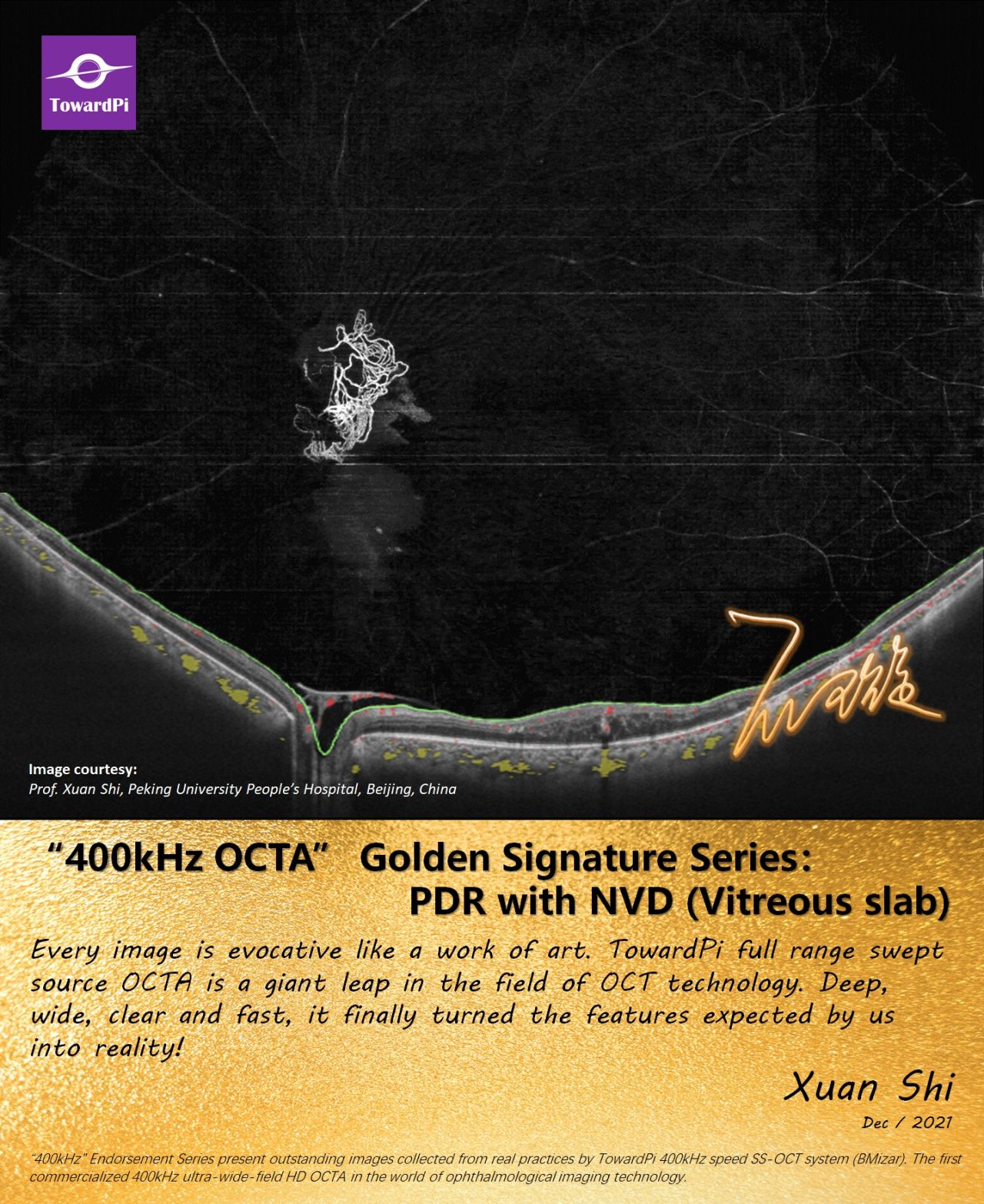
A case of retinal neovascularization in proliferative diabetic retinopathy captured by 400kHz SS-OCT/OCTA “TowardPi ultra-widefield OCT Angiography reveals retinal neovascularization and peripheral vasculature like never before, enhancing diabetic retinopathy assessment and treatment.” Golden Signature Series of TowardPi. Endorsement and courtesy: Dr. Anibal Francone (Charles Centro Oftalmológico, Argentina)
2024-10-16

How to Compare the Performance of OCT devices?

The easiest way to compare the OCT performance: SAME eye, SAME disease, SAME scan pattern, and scan at the SAME time, especially for those difficult cases (small pupil, cataract, hemorrhage, high myopia, nystagmus, RD, etc.). Illustrated figure is a case of high elevated retinal detachment with vitreous opacities.
2024-09-14

Retinal Cavernous Hemangiomas Captured with 400kHz SS-OCT/OCTA

"The combination of wide optic and swept source technology allows us to assess slow-flow moving in aneurysmal lesions and vessel alterations in wider area.”
2024-09-13

3D OCT after Artiflex IOL implantation

3D OCT after Artiflex IOL implantation, observing the position of lens in the anterior chamber and fixation on the iris. Captured with TowardPi Yalkaid Swept-Source OCT.
2024-08-20

Cyclopexy Followed Up by Full-Range SS-AS-OCT

A 58-year-old male was diagnosed with cyclodialysis and lens subluxation in his right eye. The high-resolution anterior segment OCT (AS-OCT) from 16mm x 12mm 3D AS-OCT revealed the separation of the longitudinal ciliary muscle fibers from the scleral spur (before surgery, arrow), secondary cataract and shallow anterior chamber caused by subluxation of lens. This patient underwent direct cyclopexy and IOL implantation. After surgery, the fine reattachment of the ciliary body (after surgery, arrow) and the position of IOL could be confirmed on AS-OCT.
Previous page
1
2
10

© TowardPi (Beijing) Medical Technology Ltd     京ICP备2021011388号-1   Powered by www.300.cn     View business license